

What is De Quervain’s tenosynovitis?
DeQuervain’s disease is a stenosing tenosynovitis of the first dorsal extensor compartment of the wrist. It is a condition commonly encountered in primary care and sports medicine clinics. It commonly effects women more than men and is commonly encountered in the first years of parenthood for females. (86,87)
The pathology has previously been thought secondary to inflammation causing impaired gliding of the tendons under the pulley at the thumb side of the wrist. Specifically of the abductor pollicis longus and the extensor pollicis brevis tendons. (88-90) However, recent histologic studies have shown that there are no active inflammatory cells even in patients with active disease. (88)

How is it diagnosed?
Diagnosis is often clinical with maximum tenderness and often swelling over the radial styloid and a positive Finklestein’s test. This test is done by grasping the thumb within a closed fist and then deviating the wrist ulnarly. A positive test is defined as pain at the first dorsal compartment. (86,88,89)
How is it treated?
The mainstay of treatment is conservative therapy with thumb spica wrist splinting, oral or topical anti-inflammatories and activity modification.88-91 In patients whose symptoms persist, corticosteroid injection has been the next line of therapy with up to 83% of patients experiencing relief with 1 or 2 injections.89 Other treatments that have been tried include acupuncture, massage, needle fenestration and platelet rich plasma. (91,92)
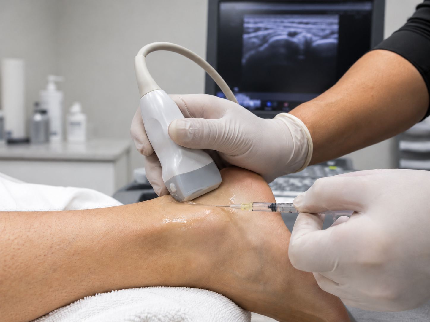
Ultrasound can be used for guided injection of the first dorsal compartment and to identify any subcompartments that may be present. (93-96) With this imaging other abnormalities can also be identified including anomalous nerve or vessel courses and persistent muscle bodies. (97-99)
Unfortunately, despite the success of conservative management and steroid injections, there are still a significant number of patients who have persistent symptoms that can be quite debilitating. (100,101) In these patients who have failed non-invasive treatments, surgical release of the first extensor compartment, including noted subcompartments, can have satisfactory results, but comes with complications associated with an invasive procedure. (96,100) More recently ultrasound-guided release of the first dorsal compartment has been described with excellent success rates. (102)
What is ultrasound guided first dorsal compartment (De Quervain’s) release?
Due to ultrasounds ability assess at risk anatomy and guide real time procedures it is reasonable to consider ultrasound-guided release of the first dorsal compartment in those patients who would rather avoid traditional open surgery or have serious medical conditions that make surgery or more extensive anesthesia more dangerous or impossible. There is precedent in using ultrasound to guided release in stenosing tenosynovitis of the flexor tendons of the fingers (trigger finger) and in carpal tunnel syndrome with good results. (69,84,103) Ultrasound-guided release has been reported in the literature with great success rates. (102)
The release itself is done by using ultrasound-guidance to direct a special needle to precisely cut the pulley through a poke hole relieving the tendons of the impingement. The tendons can then be dynamically assessed via ultrasound to assure complete release immediately following the procedure. The procedure itself is done under local anesthesia in the office and typically takes about 5 minutes to perform.
What is the recovery like?
Typically, patients can resume usual activities once the small poke hole heals (2-3 days) and are encouraged to immediately start using the wrist/hand to prevent re-scarring. Anecdotally, no pain medication outside from over-the-counter medications and ice is needed. Most patients return to complete normal function by 1-2 weeks.
What are the risks/complications?
The risks of any procedure involving a break in the skin include, infection, neurovascular or tendinous soft tissue injury and failure to provide relief. Ultrasound makes risk of iatrogenic injury to neurovascular structures and anatomic variants lower as the anatomic is viewed throughout the procedure.
References
See the full list of Dr. Pourcho's reference publications on this topic
Our Latest Stories


Book an Appointment
We're Here to Help You
Ready to take the next step in restoring your physical health? Contact us today!